Cell Cycle and Cell Division TS
Quiz Summary
0 of 13 Questions completed
Questions:
Information
You have already completed the quiz before. Hence you can not start it again.
Quiz is loading…
You must sign in or sign up to start the quiz.
You must first complete the following:
Results
Results
0 of 13 Questions answered correctly
Your time:
Time has elapsed
You have reached 0 of 0 point(s), (0)
Earned Point(s): 0 of 0, (0)
0 Essay(s) Pending (Possible Point(s): 0)
Categories
- Not categorized 0%
- 1
- 2
- 3
- 4
- 5
- 6
- 7
- 8
- 9
- 10
- 11
- 12
- 13
- Current
- Review
- Answered
- Correct
- Incorrect
- Question 1 of 13
1. Question
1 point(s)\(
\text{If a cell had } 4\ (\text{2 pairs})\ \text{chromosomes at } G_1 \text{ phase, what would be the number of chromosomes in this cell at the end of anaphase?}
\)CorrectIncorrectHint
(d)
Given:
The cell is diploid with 4 chromosomes (2 pairs) at the G₁ phase.
So, at G₁:
Chromosome number = 4 (2 from each parent — diploid)
DNA content = 4C (if we assume 1C per chromatid initially)
???? After S phase:
DNA replicates → chromatids double
Still 4 chromosomes, but each now has two sister chromatids
DNA content = 8C
???? At metaphase:
Chromosomes align at the metaphase plate
Still counted as 4 chromosomes, each with 2 sister chromatids
✂️ At anaphase:
Sister chromatids separate and move to opposite poles
Once they separate, each chromatid is considered an individual chromosome
So, chromosome number doubles
✅ Answer:
At the end of anaphase, the number of chromosomes in the cell = 8 chromosomes
???? Key Concept:
At anaphase, when sister chromatids separate, they are counted as individual chromosomes, so the chromosome number temporarily doubles.
- Question 2 of 13
2. Question
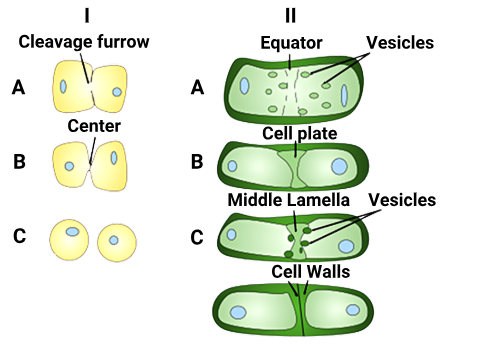
1 point(s)The diagram shows cytokinesis in animal cell [I] and in plant cell [II]. What will be correct for A, B and C in these cells?

\(
\begin{array}{|l|l|l|}
\hline & \text { Animal cell } & \text { Plant cell } \\
\hline \text { A } & \begin{array}{l}
\text { Furrow formed using } \\
\text { actin and myosin }
\end{array} & \begin{array}{l}
\text { Vesicles from Golgi move to } \\
\text { centre }
\end{array} \\
\hline \text { B } & \begin{array}{l}
\text { The furrow reaches } \\
\text { centre from periphery }
\end{array} & \begin{array}{l}
\text { Tubular structures merge to } \\
\text { form plasma membrane }
\end{array} \\
\hline \text { C } & \text { Cells pinched apart } & \begin{array}{l}
\text { Pectins deposited leading to } \\
\text { formation of middle lamella }
\end{array} \\
\hline
\end{array}
\)\(
\begin{array}{|l|l|}
\hline \text { 1. Only A and B } & \text { 2. Only A and C } \\
\hline \text { 3. Only B and C } & \text { 4. A, B and C } \\
\hline
\end{array}
\)CorrectIncorrect - Question 3 of 13
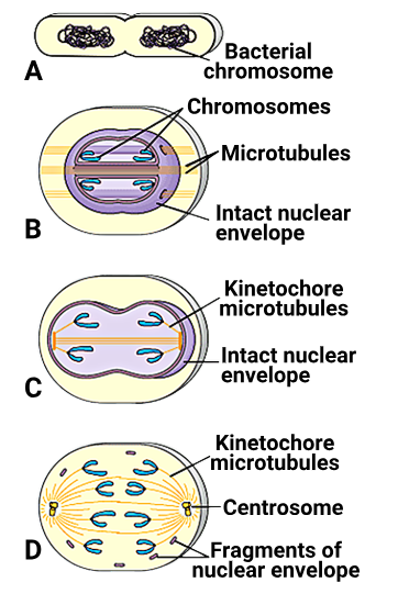
3. Question
1 point(s)The diagram given shows a hypothetical scheme of evolution of mitosis. A, B, C and D are evidences for this evolution scheme from living organisms, for example, A represents bacteria where binary fission is considered as a precursor of mitosis. Identify B, C and D:

\(
\begin{array}{|l|l|l|l|}
\hline & \text { B } & \text { C } & \text { D } \\
\hline \text { 1. } & \text { Dinoflagellates } & \text { Diatoms } & \text { Eukaryotes } \\
\hline \text { 2. } & \text { Diatoms } & \text { Euglenoids } & \text { Eukaryotes } \\
\hline \text { 3. } & \text { Yeasts } & \text { Dinoflagellates } & \text { Archaebacteria } \\
\hline \text { 4. } & \text { Diatoms } & \text { Dinoflagellates } & \text { Eukaryotes } \\
\hline
\end{array}
\)CorrectIncorrectHint
(a)
B –
Microtubules inside intact nuclear envelope
This is closed mitosis, where spindle forms inside the nucleus
Organism: ✔️ Dinoflagellates
C –
Kinetochore microtubules, intact nuclear envelope
More evolved than B, still closed mitosis
Spindle may form outside but envelope still intact
Organism: ✔️ Diatoms
D –
Fragments of nuclear envelope
Centrosomes present
Classic open mitosis of modern eukaryotes
Organism: ✔️ Eukaryotes
- Question 4 of 13
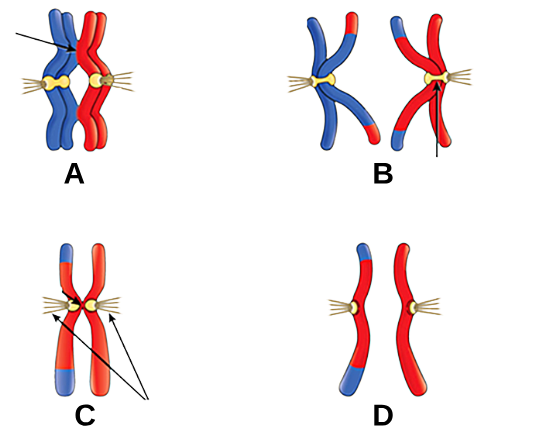
4. Question
1 point(s)A and B in the given diagram are events seen in Meiosis I and C and D are events seen in Meiosis II. Identify the correct match:

\(
\begin{array}{|c|l|}
\hline \text { A } & \text { Prophase I and arrow shows chiasmata } \\
\hline B & \begin{array}{l}
\text { Anaphase I and arrow shows sister chromatids } \\
\text { associated at centromere }
\end{array} \\
\hline C & \begin{array}{l}
\text { Prophase II and arrows show attachment of spindle } \\
\text { microtubules to kinetochore }
\end{array} \\
\hline D & \begin{array}{l}
\text { Anaphase II and the diagram shows separated sister } \\
\text { chromatids after the split of centromere }
\end{array} \\
\hline
\end{array}
\)\(
\begin{array}{|l|l|}
\hline \text { 1. Only A, B and C } & \text { 2. Only A, C and D } \\
\hline \text { 3. Only B, C and D } & \text { 4. A, B, C and D } \\
\hline
\end{array}
\)CorrectIncorrectHint
(d)
All are correct
- Question 5 of 13
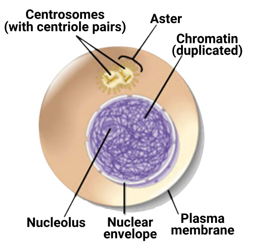
5. Question
1 point(s)At which stage of the cell cycle is the cell shown in the given diagram?

\(
\begin{array}{|l|l|}
\hline \text { 1. } G_1 & \text { 2. S } \\
\hline \text { 3. } G_2 & \text { 4. M } \\
\hline
\end{array}
\)CorrectIncorrectHint
(c) G₁ Cell grows, no DNA replication yet
S DNA is actively being replicated
G₂ DNA already replicated, centrosomes duplicate, cell prepares for mitosis ✅
M Mitosis begins, nuclear envelope breaks down ❌
- Question 6 of 13
6. Question
1 point(s)A cell at telophase stage is observed by a student in a plant brought from the field. He tells his teacher that this cell is not like other cells at telophase stage. There is no formation of cell plate and thus the cell is containing more number of chromosomes as compared to other dividing cells. This would result in
CorrectIncorrect - Question 7 of 13
7. Question
1 point(s)Anaphase promoting complex (APC) is necessary for proper mitosis of animal cells. Its main function is to
CorrectIncorrectHint
(b)
Statement 1 Prevent segregation of chromosomes❌APC does the opposite — it promotes segregation.
Statement 3 Replicate the DNA in S phase❌DNA replication is regulated by cyclins and CDKs, not APC.
Statement 4 Promote the cell from G₂ to S phase❌That’s backward — cells go from G₂ to M, and APC isn’t involved in G₂ → S
- Question 8 of 13
8. Question
1 point(s)Proteins are synthesized in preparation for mitosis during:
CorrectIncorrectHint
(c)
- Question 9 of 13
9. Question
1 point(s)The centrosome duplicates in:-
\(
\begin{array}{|l|l|}
\hline \text { 1. Early Prophase } & \text { 2. Late Prophase } \\
\hline \text { 3. S phase } & \text { 4. } \mathrm{G}_2 \text { phase } \\
\hline
\end{array}
\)CorrectIncorrectHint
(c)
- Question 10 of 13
10. Question
1 point(s)\(
\text { Cells which are not dividing are likely to be at }
\)\(
\begin{array}{|l|l|}
\hline \text { 1. } G_1 & \text { 2. } G_2 \\
\hline \text { 3. } G_0 & \text { 4. S phase } \\
\hline
\end{array}
\)CorrectIncorrectHint
(c)
- Question 11 of 13
11. Question
1 point(s)The cells in the quiescent stage \(
\left[G_0\right]
\) phase of the cell cycle:CorrectIncorrectHint
(c)
Why other options are incorrect:- 1. exit from the S phase:While cells exit the S phase (DNA synthesis) to enter the G2 phase before mitosis, G0 is not directly exiting S phase. It’s a separate phase that cells enter from G1 or sometimes directly from other phases.
- 2. continue the events of cell cycle and complete the division:The G0 phase is a pause in the cell cycle. Cells in G0 are not continuing the events of the cell cycle or completing division.
- 4. cannot ever proliferate:Cells in G0 are not currently proliferating, but they can re-enter the cell cycle and begin proliferating again when needed. This is especially true in situations like tissue repair or growth.
- Question 12 of 13
12. Question
1 point(s)Cells at the end of prophase, when viewed under the microscope, show:
CorrectIncorrectHint
(c)
During prophase, the nuclear envelope breaks down, the nucleolus disappears, and chromatin condenses into visible chromosomes. However, mitochondria, being organelles outside the nucleus, are still present throughout the cell cycle
- Question 13 of 13
13. Question
1 point(s)Liquid endosperm in coconut forms due to:
CorrectIncorrectHint
(a)
Why other options are incorrect:- 2. Failure of karyokinesis before cytokinesis:This scenario would not lead to the formation of liquid endosperm. If karyokinesis failed, there would be no nuclei to divide, and thus no endosperm would develop.
- 3. Disruption of spindle fibers at metaphase:While spindle fiber disruption can lead to abnormal cell division, it wouldn’t specifically result in the formation of liquid endosperm. Disruption at metaphase would more likely cause aneuploidy (abnormal chromosome number) in the daughter cells.
- 4. Continued DNA replication during cytokinesis:DNA replication occurs during the S phase of the cell cycle, before mitosis, not during cytokinesis. Cytokinesis is the division of the cytoplasm, not the replication of DNA.