2.9 Exercise Problems
Q1. Fill in the blanks:
(a) Humans reproduce _______ (asexually/sexually).
(b) Humans are_________ (oviparous, viviparous, ovoviviparous).
(c) Fertilization is in humans ______ (external/internal).
(d) Male and female gametes are __________ (diploid/haploid).
(e) Zygote is __________ (diploid/haploid).
(f) The process of release of ovum from a mature follicle is called __________
(g) Ovulation is induced by a hormone called __________
(h) The fusion of male and female gametes is called __________
(i) Fertilization takes place in __________
(j) Zygote divides to form _________ which is implanted in uterus.
(k) The structure which provides vascular connection between foetus and uterus is called ______
Answer: (a) sexually
(b) viviparous
(c) internal
(d) haploid
(e) diploid
(f) ovulation
(g) LH (Luteinizing hormone)
(h) fertilization
(i) ampullary-isthmic junction (fallopian tube)
(j) blastocyst
(k) placenta (Umbilical cord)
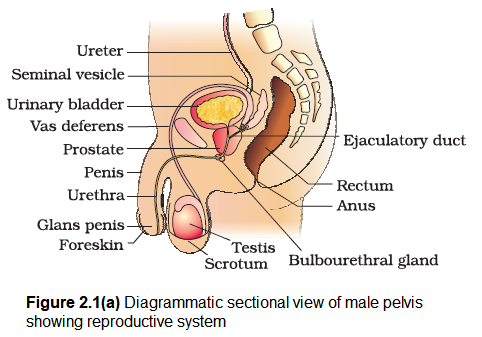
Q2. Draw a labelled diagram of male reproductive system.
Answer:
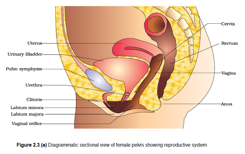
Q3. Draw a labelled diagram of female reproductive system.
Answer:
Q4. Write two major functions each of testis and ovary.
Answer: Testes are components of both the reproductive system (being gonads) and the endocrine system (being endocrine glands). The respective functions of the testes are – producing sperm (spermatozoa) by the process of spermatogenesis and producing male sex hormones, of which testosterone is the best known. Testosterone stimulates development of testes and of male secondary sexual characteristics.
The ovaries have two major functions. One is the production of eggs or ova, and the second is the production of hormones or chemicals which regulate menstruation and other aspects of health and wellbeing, including sexual well-being. Estrogen and progesterone are the most important hormones which serve many functions like, they induce and maintain the physical changes during puberty and the secondary sex characteristics and they support maturation of the uterine endometrium in preparation for implantation for a fertilised egg, etc.
Q5. Describe the structure of a seminiferous tubule.
Answer: The seminiferous tubule is a structural unit in the adult testis. The seminiferous tubules are situated in testicular lobules. Seminiferous tubule consists of two types of cells . Sertoli or supporting cells & spermatogenic cells, Sertoli cells , are elongated and pyramidal & partially envelop the spermatogenic cells. The cells provide nourishment to the developing spermatogenic cells. Spermatogenic cells are stacked in 48 layers. These cells divide several times & differentiate to produce spermatozoa. Between seminiferous tubules lie the interstitial cells or Leydig cells which produces testosterone hormone.
Q6. What is spermatogenesis? Briefly describe the process of spermatogenesis.
Answer: Spermatogenesis is the process of producing sperms with half the number of chromosomes (haploid) as somatic cells. It occurs in seminiferous tubules. Sperm production begins at puberty continues throughout life with several hundred million sperms being produced each day. Once sperm are formed they move into the epididymis, where they mature and are stored. During spermatogenesis one spermatogonium produces 4 sperms. Spermatogenesis completes through the following phases – multiplicative phase, growth phase, maturation phase & spermiogenesis. In multiplicative phase the sperm mother cells divide by mitosis & produce spermatogonia. The spermatogonia grow in size to form large primary spermatocytes by getting nourishment from sertoli cells in growth phase. Maturation phase involves meiosis I in which primary spermatocytes divide to produce secondary spermatocyte and meiosis II which produces spermatids. Thus each primary spermatocyte gives rise to four haploid spermatids. Spermiogenesis or spermatogenesis is process of formation of flagellated spermatozoa from spermatids. Spermiogenesis begins in the seminiferous tubules but usually completed in epididymis.
Q7. Name the hormones involved in regulation of spermatogenesis.
Answer: The hormones involved in regulation of spermatogenesis are \(\mathrm{GnRH}, \mathrm{LH}, \mathrm{FSH}\) and androgens. Spermatogenesis starts at the age of puberty due to significant increase in the secretion of gonadotropin releasing hormone (GnRH). The increased levels of GnRH then acts at the anterior pituitary gland and stimulates secretion of two gonadotropins – luteinising hormone (LH) and follicle stimulating hormone (FSH). LH acts at the Leydig cells and stimulates synthesis and secretion of androgens. Androgens, in turn, stimulate the process of spermatogenesis. FSH acts on the Sertoli cells and stimulates secretion of some factors which help in the process of spermiogenesis.
Q.8. Define spermiogenesis and spermiation.
Answer: Spermiogenesis is the process of transformation of spermatids into mature flagellated spermatozoa (sperms). Spermiation is the process of release of mature spermatozoa. In this spermatozoa are shed into the lumen of seminiferous tubule for transport.
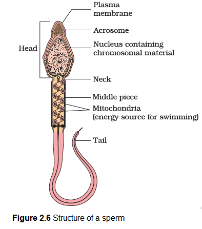
Q9. Draw a labelled diagram of sperm.
Answer:
Q10. What are the major components of seminal plasma?
Answer: Seminal plasma is the fluid in which sperm is ejaculated. Major components of seminal plasma are secretions from seminal vesicles, prostrate and bulbourethral gland and sperms from testis . It is rich in fructose and contains enzymes, citric acid, hormones like prostaglandins, calcium and clotting proteins.
Q11. What are the major functions of male accessory ducts and glands?
Answer: Male accessory ducts include rete testis, vasa efferentia, epididymis and vas deferens. These ducts store and transport sperms from the testis to the outside through urethra. The male accessory glands include paired seminal vesicles, a prostate and paired bulbourethral glands. Secretions of these glands constitute the seminal plasma which is rich in fructose, calcium and certain enzymes. The secretions of bulbourethral glands also helps in the lubrication of the penis.
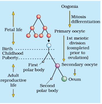
Q12. What is oogenesis? Give a brief account of oogenesis.
Answer: The process of formation of a mature female gamete (ovum) is called oogenesis. It occurs in the ovaries of female reproductive system. Oogenesis is a discontinuous process it begins before birth, stops in midprocess & only resumes after menarch. It occurs in three phases : Multiplicative phase (formation of oogonia mitotically from the primary germ cells), Growth phase (growth of oogonia into primary oocyte) & Maturation phase (formation of mature ova from primary oocyte through meiosis). Maturation phase produces two haploid cells – Larger one called secondary oocyte & the smaller one called polar bodies (1st polar body). Meiosis II of secondary oocyte results in the formation of functional egg or ovum and a second polar body: The first polar body may also divide to form two polar bodies of equal sizes which do not take part in reproduction & ultimately degenerates. First maturation division may be completed in the ovaries just prior to ovulation but second one (Final) is completed outside the ovary after fertilization. Secondary oocyte is female gamete in which the 1st meiotic division is completed & second meiotic division (Metaphase stage) has begin. The egg is released at secondary oocyte stage under the effect of LH.
Q13. Draw a labelled diagram of a section through ovary.
Answer:
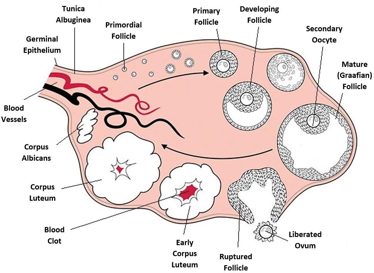
Q14. Draw a labelled diagram of a Graafian follicle.
Answer:
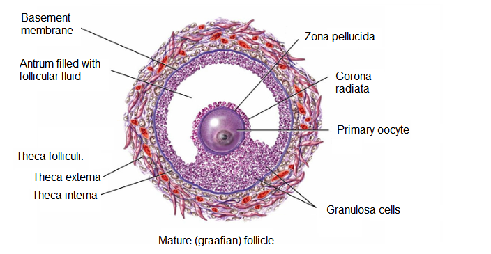
Q15. Name the functions of the following:
(a) Corpus luteum
(b) Endometrium
(c) Acrosome
(d) Sperm tail
(e) Fimbriae
Answer: (a) Corpus luteum: The corpus luteum secretes large amounts of progesterone which is essential for maintenance of the endometrium.
(b) Endometrium is necessary for implantation of the fertilized ovum and other events of pregnancy.
(c) The acrosome is filled with enzymes that help during fertilization of the ovum.
(d) Sperm tail: Tail facilitates sperm motility which is essential for fertilization.
(e) Fimbriae: Fimbriae help in collection of the ovum after ovulation.
Q16. Identify True/False statements. Correct each false statement to make it true.
(a) Androgens are produced by Sertoli cells. (True/False)
(b) Spermatozoa get nutrition from sertoli cells. (True/False)
(c) Leydig cells are found in ovary. (True/ False)
(d) Leydig cells synthesize androgens. (True/ False)
(e) Oogenesis takes place in corpus luteum. (True/False)
(i) Menstrual cycle ceases during pregnancy. (True/False)
(g) Presence or absence of hymen is not a reliable indicator of virginity or sexual – experience. (True/False)
Answer: (a) False, Androgens or male sex hormones (e.g., testosterone) are secreted by Leydig cells.
(b) True.
(c) False, Leydig cells are found in testis.
(d) True.
(e) False, Oogenesis takes place in ovary.
(f) True.
(g) True.
Q17. What is menstrual cycle? Which hormones regulate menstrual cycle?
Answer: Menstrual cycle is the cyclic change( in the reproductive tract of primate female) . This period is marked by a characteristic event repeated almost every month ( 28 days with minor variation) in the form of a menstrual flow (i.e. shedding of the endometrium of the uterus with bleeding. It may be temporarily stopped only in pregnancy.
The hormones that regulates menstrual cycles are
(i) FSH (Follicle stimulating hormone),
(ii) LH (Luteinizing hormone),
(iii) Oestrogens (estrogens),
(iv) Progesterone.
Q18. What is parturition ? Which hormones are involved in induction of parturition?
Answer: Parturition (or labour) means child birth. Parturition is the sequence of actions by which a baby and the after birth (placenta) are expelled from the uterus at childbirth. The process usually starts spontaneously about 280 days after conception, but it may be started by artificial means. The process of parturition is induced by a complex neuroendocrine mechanisms involving cortisol, estrogen and oxytocin.
Q19. In our society the women are often blamed for giving birth to daughters. Can you explain why this is not correct?
Answer: The sex chromosome pattern in the human females is \(X X\) and that of male is \(X Y\). Therefore, all the haploid female gametes (ova) have the sex chromosome \(X\), however, the haploid male gametes have either \(X\) or \(Y\). Thus \(50 \%\) of sperms carry the \(X\)-chromosome while the other \(50 \%\) carry the \(Y\)-chromosome. After fusion of the male and female gametes, the zygote carries either \(X X\) or \(X Y\) depending upon whether the sperm carrying \(X\) or \(Y\) fertilizes the ovum. The zygote carrying \(X X\) would be a female baby and \(X Y\) would be a male baby. That is why it is correct to say that the sex of the baby is determined by the father.
Q20. How many eggs are released by a human ovary in a month? How many eggs do you think would have been released if the mother gave birth to identical twins? Would your answer change if the twins born were fraternal?
Answer: One egg is released by human ovary in a month. Identical twins: Identical twins are formed when a single fertilized egg splits into two genetically identical parts. The twins share the same DNA set, thus they may share many similar attributes. However, since physical appearance is influenced by environmental factors and not just genetics, identical twins can actually look very different.
Fraternal twins: These twins are formed when two fertilized eggs are formed. The twins share the different DNA sets, thus they may share different attributes (dizygotic embryo).
Q21. How many eggs do you think were released by the ovary of a female dog which gave birth to 6 puppies?
Answer: Since dogs have multiple births, several eggs mature and are released at the same time. If fertilised, the egg will implant on the uterine wall. Dogs bear their litters roughly 9 weeks after fertilisation, although the length of gestation can vary from 56 to 72 days. An average litter consists of about six puppies, though this number may vary widely based on the breed of dog. On this basis 6 eggs were released by the ovary of a female dog which gave birth to 6 puppies.
Exemplar Section
VERY SHORT ANSWER TYPE QUESTIONS
Q1. Given below are the events in human reproduction. Write them in correct sequential order.
Insemination, gametogenesis, fertilisation, parturition, gestation, implantation
Answer: Gametogenesis -> Insemination -> Fertilisation ->Implantation -> Gestation -> Parturition
Q2. The path of sperm transport is given below. Provide the missing steps in blank boxes.

Answer:

Q3. What is the role of cervix in the human female reproductive system?
Answer: Cervix helps in regulating the passage of sperms into the uterus and forms the birth canal to facilitate parturition.
Q4. Why are menstrual cycles absent during pregnancy.
Answer: The high levels of progesterone and estrogens during pregnancy suppress the gonadotropins which is required for the development of new follicles. Therefore, a new cycle cannot be initiated.
Q5. Female reproductive organs and associated functions are given below in column A and B. Fill the blank boxes.
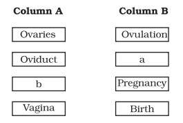
Answer: a – Fertilisation, b – Uterus
Q6. From where the parturition signals arise-mother or foetus? Mention the main hormone involved in parturition.
Answer: The signals for parturition originate from the fully developed foetus and the placenta which induce mild uterine contractions called foetal ejection reflex. This triggers release of oxytocin from the maternal pituitary. The main hormone involved in parturition is oxytocin.
Q7. What is the significance of epididymis in male fertility?
Answer: In the epididymis sperms undergo physiological maturation, acquiring increased motility and fertilizing capacity.
Q8. Give the names and functions of the hormones involved in the process of spermatogenesis. Write the names of the endocrine glands from where they are released.
Answer: Spermatogenesis starts at the age of puberty due to significant increase in the secretion of gonadotropin releasing hormone (GnRH). This is a hypothalamic hormone.
- The increased levels of GnRH then acts at the anterior pituitary gland and stimulates secretion of two gonadotropins leutinising hormone (LH) and follicle stimulating hormone (FSH).
- LH acts at the Leydig cells and stimulates synthesis and secretion of androgens. Androgens, in turn, stimulate the process of spermatogenesis.
- FSH acts on the Sertoli cells and stimulates secretion of some factors which help in the process of spermiogenesis.
Q9. The mother germ cells are transformed into a mature follicle through series of steps. Provide the missing steps in the blank boxes.

Answer: a- Primary oocyte, b-Tertiary follicle, c- Secondary follicle
Q10. During reproduction, the chromosome number (2n) reduces to half (n) in the gametes and again the original number (2n) is restored in the offspring, What are the processes through which these events take place?
Answer: Halving of chromosomal number takes place during gametogenesis and regaining the 2n number occur as a result of fertilisation.
Q11. What is the difference between a primary oocyte and a secondary oocyte?
Answer:
\(\begin{array}{|c|l|c|l|}
\hline & \text { Primary oocytes } & & \text { Secondary oocytes } \\
\hline \text { 1. } & \text { Diploid } & 1 . & \text { Haploid } \\
\hline 2 . & \begin{array}{l}
\text { Formed from oogonia } \\
\end{array} & 2 . & \begin{array}{l}
\text { Developed from the primary } \\
\text { ocyte through meiosis I }
\end{array} \\
\hline \text { 3. } & \text { Found in primary follicles } & 3 . & \begin{array}{l}
\text { Found in the matured Graafian } \\
\text { follicle. }
\end{array} \\
\hline
\end{array}
\)
Q12. What is the significance of ampullary–isthmic junction in the female reproductive tract?
Answer: In mammals, fertilization takes place in the ampullary-isthmic junction.
Q13. How does zona pellucida of ovum help in preventing polyspermy?
Answer: Polyspermy means an egg has been fertilized by more than one sperms. The zona pellucida is modified by proteases. The proteases destroy the protein link between the cell membrane and the vitelline membrane, remove any receptors that other sperm have bound to, and help to form the fertilization membrane that prevents polyspermy.
Q14. Mention the importance of LH surge during menstrual cycle.
Answer: LH surge is essential for the events leading to ovulation.
Q15. Which type of cell division forms spermatids from the secondary spermatocytes?
Answer: Second meiotic division
SHORT ANSWER TYPE QUESTIONS
Q1. A human female experiences two major changes, menarche and menopause during her life. Mention the significance of both the events.
Answer: Menarche represents the beginning of menstrual cycle which is an indication of attainment of sexual maturity. Menopause, on the other hand, refers to the cessation of menstruation which in turn means stoppage of gamete production, i.e., it marks the end of reproductive/ fertile life of the female.
Q2. a. How many spermatozoa are formed from one secondary spermatocyte?
b. Where does the first cleavage division of zygote take place?
Answer: (a) Two spermatozoa are formed from one secondary spermatocyte.
(b) First cleavage division of zygote take place in fallopian tube or oviduct.
Q3. Corpus luteum in pregnancy has a long life. However, if fertilisation does not take place, it remains active only for 10-12 days. Explain.
Answer: This is because of a neural signal given by the maternal endometrium to its hypothalamus in presence of a zygote to sustain the gonadotropin (LH) secretion, so as to maintain the corpus luteum as long as the embryo remains there. In the absence of a zygote, therefore, the corpus luteum cannot be maintained longer.
Q4. What is foetal ejection reflex? Explain how it leads to parturition?
Answer: Vigorous contraction of the uterus at the end of the pregnancy causes expulsion/delivery of the foetus. This process of delivery of the foetus (childbirth) is called parturition.
- Parturition is induced by a complex neuroendocrine mechanism. The signals for parturition originate from the fully developed foetus and the placenta which induce mild uterine contractions called foetal ejection reflex. This triggers release of oxytocin from the maternal pituitary. Oxytocin acts on the uterine muscle and causes stronger uterine contractions, which in turn stimulates further secretion of oxytocin.
- The stimulatory reflex between the uterine contraction and oxytocin secretion continues resulting in stronger and stronger contractions. This leads to expulsion of the baby out of the uterus through the birth canal- parturition. Soon after the infant is delivered, the placenta is also expelled out of the uterus.
Q5. Except endocrine function, what are the other functions of placenta.
Answer: Placenta facilitates the supply of oxygen and nutrients to the embryo. It also removes \(\mathrm{CO}_2\) and excretory wastes produced by embryo.
Q6. Why doctors recommend breast feeding during initial period of infant growth?
Answer: The mammary glands of the female undergo differentiation during pregnancy and starts producing milk towards the end of pregnancy by the process called lactation. The milk produced during the initial few days of lactation is called colostrum which contains several antibodies absolutely essential to develop resistance for the new born babies. Breast-feeding during the initial period of infant growth is recommended by doctors for bringing up a healthy baby.
Q7. What are the events that take place in the ovary and uterus during follicular phase of the menstrual cycle.
Answer: 1. The primary follicle .grows and becomes fully mature graafian follicles.
2. Secretion of estrogen hormone.
3. Endometrium of uterus regenerates through proliferation.
Q8. Given below is a flow chart showing ovarian changes during menstrual cycle. Fill in the spaces giving the name of the hormones responsible for the events shown.
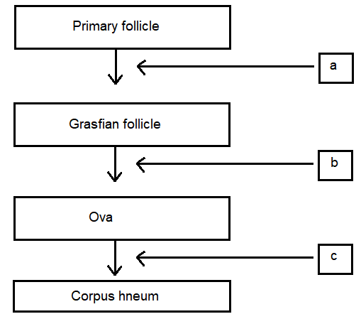
Answer: a – FSH and LH, b – LH, c – Progesterone
Q9. Give a schematic labelled diagram to represent oogenesis (without descriptions)
Answer:
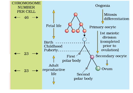
Q10. What are the changes in the oogonia during the transition of a primary follicle to Graafian follicle?
Answer: Oogenesis is initiated during the embryonic development stage when a couple of million gamete mother cells (oogonia) are formed within each fetal ovary (about third month of foetal ovary), no more oogonia are formed and added after birth.
- These cells start division and enter into prophase-l of the meiotic division and get temporarily arrested at that stage, called primary oocytes. Each primary oocyte then gets surrounded by a layer of granulosa cells and then called the primary follicle. \(1^{\circ}\) follicle contains \(1^{\circ}\) oocyte. A large number of these follicles degenerate during the phase from birth to puberty.
- The primary follicles get surrounded by more layers of granulosa cells and a new theca and called secondary follicles. The theca layer is organised into an inner theca interna and an outer theca externa. Theca interna secretes estrogen hormone. The secondary follicle soon transforms into a tertiary follicle which is characterised by a fluid filled cavity called antrum.
- It is important to draw your attention that it is at this stage that the primary oocyte within the tertiary follicle grows in size and completes its first meiotic division. It is an unequal division resulting in the formation of a large haploid secondary oocyte and a tiny first polar body. The tertiary follicle further changes into the mature follicle or Graafian follicle.
LONG ANSWER QUESTIONS
Q1. What role does pituitary gonadotropins play during follicular and ovulatory phases of menstrual cycle? Explain the shifts in steroidal
secretions.
Answer: Menstrual cycle is regulated by hypothalamus through the pituitary gland. At the end of menstrual phase, the pituitary FSH gradually increases resulting in follicular development within the ovaries. As the follicles mature, Estrogen secretion increases resulting in a surge in (FSHe and LH).The surge of LH responsible for ovulation. LH also induces luteinisation. This leading to the formation of corpus luteum. Corpus luteum secretes progesterone and some estrogen which help in maintaining the uterine endometrium for implantation.
Q2. Meiotic division during oogenesis is different from that in spermatogenesis. Explain how and why?
Answer: Unequal cytoplasmic division of the oocyte is to ensure the retention of bulk of cytoplasm in one cell, instead of sharing with two. It has to provide nourishment for the developing embryo during early stages, so it is essential to retain as much cytoplasmic materials it could in a single daughter cell.
Q3. The zygote passes through several developmental stages till implantation, Describe each stage briefly with suitable diagrams.
Answer: The mitotic division starts as the zygote moves through the isthmus of the oviduct called cleavage towards the uterus and forms 2, 4, 8, 16 daughter cells called blastomeres. The embryo with 8 to 16 blastomeres is called a morula. The morula continues to divide and transforms into blastocysts, it moves further into the uterus.
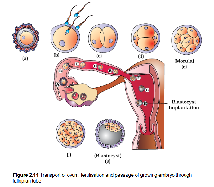
- The blastomeres in the blastocyst are arranged into an outer layer called trophoblast and an inner group of cells attached to trophoblast called the inner cell mass. The trophoblast layer then gets attached to the endometrium and the inner cell mass gets differentiated as the embryo.
- After attachment, the uterine cells divide rapidly and covers the blastocyst. As a result the blastocyst becomes embedded in the endometrium of the uterus. This is called implantation and it leads to pregnancy.
Q4. Draw a neat diagram of the female reproductive system and label the parts associated with the following (a) production of gamete, (b) site of fertilisation (c) site of implantation and, (d) birth canal.
Answer:
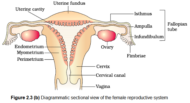
\left.\begin{array}{l}
\text { Cervical canal } \\
\text { Vagina }
\end{array}\right] \begin{aligned}
& \text { Birth } \\
& \text { canal }
\end{aligned}
\)
Q5. With a suitable diagram, describe the organisation of mammary gland.
Answer: A functional mammary gland is characteristic of all female mammals. The mammary glands are paired structures (breasts) that contain glandular tissue and variable amount of fat. The glandular tissue of each breast is divided into 15-20 mammary lobes containing clusters of cells called alveoli. The cells of alveoli secrete milk, which is stored in the cavities (lumens) of alveoli.
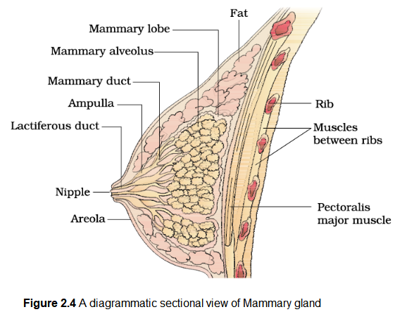
The alveoli open into mammary tubules. The tubules of each lobe join to form a mammary duct. Several mammary ducts join to form a wider mammary ampulla which is connected to lactiferous ducts through which milk is sucked out.