2.3 Gametogenesis
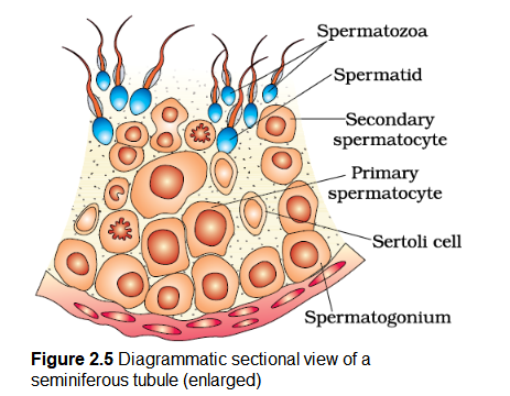
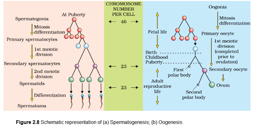
The primary sex organs – the testis in the males and the ovaries in the females-produce gametes, i.e, sperms and ovum, respectively, by the process called gametogenesis. In testis, the immature male germ cells (spermatogonia) produce sperms by spermatogenesis that begins at puberty. The spermatogonia (sing. spermatogonium) present on the inside wall of seminiferous tubules multiply by mitotic division and increase in numbers. Each spermatogonium is diploid and contains 46 chromosomes. Some of the spermatogonia called primary spermatocytes periodically undergo meiosis. A primary spermatocyte completes the first meiotic division (reduction division) leading to formation of two equal, haploid cells called secondary spermatocytes, which have only 23 chromosomes each. The secondary spermatocytes undergo the second meiotic division to produce four equal, haploid spermatids (Figure 2.5). What would be the number of chromosome in the spermatids? The spermatids are transformed into spermatozoa (sperms) by the process called spermiogenesis. After spermiogenesis, sperm heads become embedded in the Sertoli cells, and are finally released from the seminiferous tubules by the process called spermiation.

Spermatogenesis starts at the age of puberty due to significant increase in the secretion of gonadotropin releasing hormone (GnRH). This, if you recall, is a hypothalamic hormone. The increased levels of GnRH then acts at the anterior pituitary gland and stimulates secretion of two gonadotropins – luteinising hormone (LH) and follicle stimulating hormone (FSH). LH acts at the Leydig cells and stimulates synthesis and secretion of androgens. Androgens, in turn, stimulate the process of spermatogenesis. FSH acts on the Sertoli cells and stimulates secretion of some factors which help in the process of spermiogenesis.
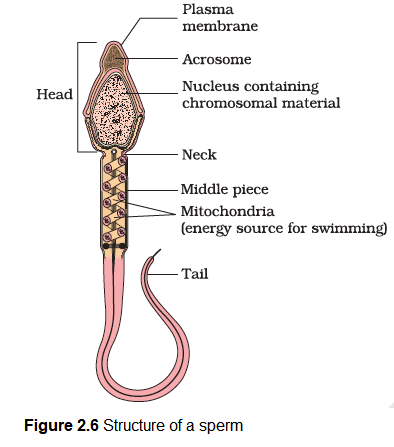
Let us examine the structure of a sperm. It is a microscopic structure composed of a head, neck, a middle piece and a tail (Figure 2.6). A plasma membrane envelops the whole body of sperm. The sperm head contains an elongated haploid nucleus, the anterior portion of which is covered by a cap-like structure, acrosome. The acrosome is filled with enzymes that help fertilisation of the ovum. The middle piece possesses numerous mitochondria, which produce energy for the movement of tail that facilitate sperm motility essential for fertilisation. The human male ejaculates about 200 to 300 million sperms during a coitus of which, for normal fertility, at least 60 percent sperms must have normal shape and size and at least 40 per cent of them must show vigorous motility.

Sperms released from the seminiferous tubules, are transported by the accessory ducts. Secretions of epididymis, vas deferens, seminal vesicle and prostate are essential for maturation and motility of sperms. The seminal plasma along with the sperms constitute the semen. The functions of male sex accessory ducts and glands are maintained by the testicular hormones (androgens).
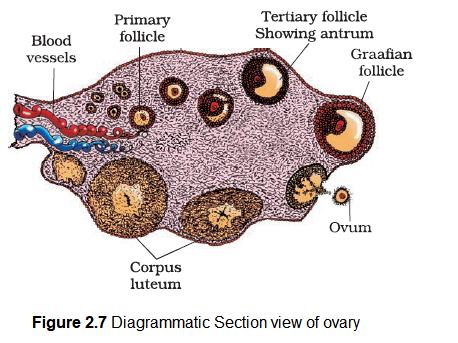
The process of formation of a mature female gamete is called oogenesis which is markedly different from spermatogenesis. Oogenesis is initiated during the embryonic development stage when a couple of million gamete mother cells (oogonia) are formed within each fetal ovary; no more oogonia are formed and added after birth. These cells start division and enter into prophase-I of the meiotic division and get temporarily arrested at that stage, called primary oocytes. Each primary oocyte then gets surrounded by a layer of granulosa cells and is called the primary follicle (Figure 2.7). A large number of these follicles degenerate during the phase from birth to puberty. Therefore, at puberty only 60,000-80,000 primary follicles are left in each ovary. The primary follicles get surrounded by more layers of granulosa cells and a new theca and are called secondary follicles.

The secondary follicle soon transforms into a tertiary follicle which is characterised by a fluid filled cavity called antrum. The theca layer is organised into an inner theca interna and an outer theca externa. It is important to draw your attention that it is at this stage that the primary oocyte within the tertiary follicle grows in size and completes its first meiotic division. It is an unequal division resulting in the formation of a large haploid secondary oocyte and a tiny first polar body (Figure 2.8b). The secondary oocyte retains bulk of the nutrient rich cytoplasm of the primary oocyte. Can you think of any advantage for this? Does the first polar body born out of first meiotic division divide further or degenerate? At present we are not very certain about this. The tertiary follicle further changes into the mature follicle or Graafian follicle (Figure 2.7). The secondary oocyte forms a new membrane called zona pellucida surrounding it. The Graafian follicle now ruptures to release the secondary oocyte (ovum) from the ovary by the process called ovulation. Can you identify major differences between spermatogenesis and oogenesis? A diagrammatic representation of spermatogenesis and oogenesis is given below (Figure 2.8).
